Home » Without Label » Back Muscle Diagram : Upper Back Pain Anatomy Of The Back The Pain Center Pain Management Care - The part of the nerve that emerges out of the spine is called the nerve root.
Back Muscle Diagram : Upper Back Pain Anatomy Of The Back The Pain Center Pain Management Care - The part of the nerve that emerges out of the spine is called the nerve root.
Back Muscle Diagram : Upper Back Pain Anatomy Of The Back The Pain Center Pain Management Care - The part of the nerve that emerges out of the spine is called the nerve root.. We hope this picture anatomy of back muscles diagram can help you study and research. When back development is the goal, stick to one of these variations. The part of the nerve that emerges out of the spine is called the nerve root. Stand behind the barbell with your feet shoulder. Likewise, there are muscles in other parts of the body that help support and move the spine.
The muscles on each side form a trapezoid shape. While muscles like the gluteals (in the thighs) are used any time we walk or climb a step, deep back muscles and abdominal muscles are usually not actively engaged during everyday activity. The muscles of the back can be arranged into 3 categories based on their location: We hope this picture anatomy of back muscles diagram can help you study and research. Posted on may 12, 2015 by admin.
Muscles Move And Support The Spine from cloud2.spineuniverse.com Working the lower back, erector spinae muscles, and hamstrings, a barbell deadlift requires back strength to effectively complete. It contains the osteology, arthrology and myology of the spine and back. These structures work together to support the body, enable a range of movements, and send messages from the. Creatine is now proving to be one of the most potent muscle growth accelerators giving excellent muscle mass increase and phenomenal strength increases order yours today. Others, like sumo deadlifts, have been shown in emg studies—and in the trenches—to focus more on other muscle groups than the back. When back development is the goal, stick to one of these variations. Muscle anatomy crossword answer 12 photos of the muscle anatomy crossword answer muscle anatomy crossword answer key biology corner, muscle anatomy crossword answers biology corner, muscle anatomy crossword answers key, muscle anatomy crossword puzzle answers biology corner, muscle anatomy. To learn more about the anatomy of the spine, watch this video.
Chronic back pain map this tool recommended for:
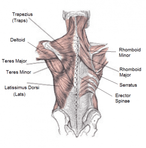
To learn more about the anatomy of the spine, watch this video. Likewise, there are muscles in other parts of the body that help support and move the spine. Below you'll see diagrams along with the names of the back muscles that may be the cause of your pain. It is the most superficial of all the back muscles. The trapezius and latissimus dorsi muscles connect the upper limb to the vertebral column. Pain log more pain mapping tools The fibres attach to the clavicle, acromion and the scapula spine. We hope this picture anatomy of back muscles diagram can help you study and research. The part of the nerve that emerges out of the spine is called the nerve root. The deltoid, teres major, teres minor, infraspinatus, supraspinatus (not shown) and subscapularis muscles (not shown) all extend from the scapula to the humerus and act on the shoulder joint. Posted on may 12, 2015 by admin. Nerves in your lower back. The back has a total of 40 muscles.
This human anatomy module is composed of diagrams, illustrations and 3d views of the back, cervical, thoracic and lumbar spinal areas as well as the various vertebrae. Five pairs of lumbar spinal nerves labeled l1 to l5 branch off your spinal cord and exit through small holes between the vertebrae. The trapezius and latissimus dorsi muscles connect the upper limb to the vertebral column. Deep back muscles diagram the superficial layer contains the splenius cervicis and splenius capitis muscles. Back to tracking tools main page.
Labeled Anatomy Chart Of Male Lower Back Muscles On White Background Stock Photo Alamy from c8.alamy.com See back muscles and low back pain. Chronic back pain map this tool recommended for: Likewise, there are muscles in other parts of the body that help support and move the spine. Human anatomy diagrams show internal organs, cells, systems, conditions, symptoms and sickness information and/or tips for healthy living. This is a diagram of the larger and more surface muscles of the low back. Superficial, intermediate, deep and deepest layers.these muscles lie on each side of the vertebral column, deep to the thoracolumbar fascia they span the entire length of the vertebral column, extending from the cranium to the pelvis What is the origin and insertion of the rhomboid minor and major muscle? This diagram depicts muscles in back diagram.
The back has a total of 40 muscles.
The fibres attach to the clavicle, acromion and the scapula spine. What is the origin and insertion of the rhomboid minor and major muscle? It is the most superficial of all the back muscles. Below you'll see diagrams along with the names of the back muscles that may be the cause of your pain. Anatomy of the spine and back spine muscles diagram. We think this is the most useful anatomy picture that you need. A strained muscle in your lower back can be quite painful. These structures work together to support the body, enable a range of movements, and send messages from the. Muscle anatomy crossword answer 12 photos of the muscle anatomy crossword answer muscle anatomy crossword answer key biology corner, muscle anatomy crossword answers biology corner, muscle anatomy crossword answers key, muscle anatomy crossword puzzle answers biology corner, muscle anatomy. The pelvis at the bottom of the back and the shoulders at the top of the back give the back. Deep back muscles diagram the superficial layer contains the splenius cervicis and splenius capitis muscles. It is particularly interesting for physiotherapists. Your back hurting more when you move, less when you stay still;
Most of the time, back muscle pain is diagnosed then treated with little more than a prescription of rest, painkillers and muscle relaxants. Muscle strain is often the cause of back pain from heavy lifting or vigorous exercise. The intermediate layer contains the erector spinae muscles, whose many functions include the extension and lateral flexion of the spine, head and neck. Back to tracking tools main page. Posted on may 12, 2015 by admin.
The Complete Guide To Training Your Back from 9to5strength.com Below you'll see diagrams along with the names of the back muscles that may be the cause of your pain. Nerves in your lower back. It is opposite from the chest, and the vertebral column runs down the back. Anatomy of the spine and back spine muscles diagram. They extend and rotate the head and neck. When back development is the goal, stick to one of these variations. Pain log more pain mapping tools The deltoid, teres major, teres minor, infraspinatus, supraspinatus (not shown) and subscapularis muscles (not shown) all extend from the scapula to the humerus and act on the shoulder joint.
Back to tracking tools main page.
How many muscles are in the back? Muscles labeled front and back 12 photos of the muscles labeled front and back muscle diagram labeled front and back, muscle system labelling (front and back), muscular system labeled front and back, human muscles, muscle diagram labeled front and back, muscle system labelling (front and back), muscular system labeled front and back. Five pairs of lumbar spinal nerves labeled l1 to l5 branch off your spinal cord and exit through small holes between the vertebrae. Your back hurting more when you move, less when you stay still; People with back pain people who experience headaches printing for use during doctor visits to communicate information about your symptoms quickly tracking your progress over time related tools: Nerves in your lower back. This diagram depicts muscles in back diagram. The pelvis at the bottom of the back and the shoulders at the top of the back give the back. Both the deltoid and the trapezius are firmly attached to the spine of the scapula. In this image, you will find 1st cervical vertebrae, atlus, cervical plexus, 7th cervical vertebrae, 1st thoracic vertebrae, brachial plexus, spinal dura mater, filaments of spinal nerve roots, 12th thoracic vertebra, 1st lumber vertebra, iliohypogastric nerve, ilioinguinal nerve, lumbar. It contains the osteology, arthrology and myology of the spine and back. For example, some muscles located in the chest also help move the shoulders. These structures work together to support the body, enable a range of movements, and send messages from the.